Oral Presentation Crystal36-AXAA Conference 2026
Revealing cement hydration transformations through long-term in-situ XRD and complementary techniques (143107)
Globally, Portland cement (PC) is one of the most widely consumed construction materials, with annual production reaching approximately 4.6 billion tonnes. Forecasts project demand in 2050 to range between 3.7-5.5 billion tonnes [1]. However, PC production has significant environmental impacts, contributing approximately 8% of global CO₂ emissions [2]. Consequently, there is a strong drive to develop more sustainable alternatives, including blended cement systems incorporating supplementary cementitious materials (SCMs) and other cementitious formulations [3].
Cement hydration is a complex, long-term process, with phase evolution continuing for months after initial mixing. This presents challenges for extended investigations, particularly in multi-user characterisation facilities. In this study, we present a method for in-situ X-ray diffraction (XRD) to investigate the hydration of Ordinary Portland Cement (OPC). Complementary microanalysis was also conducted using elemental X-ray mapping (XRM) and micro-computed tomography (micro-CT).
XRD measurements were performed using a Bruker D8 Advance diffractometer in Bragg–Brentano geometry, equipped with a Cu X-ray source operating at 40 kV and 40 mA, and a LynxEye XE-T detector. OPC paste was prepared and mounted in a custom airtight holder, sealed with Kapton foil. In-situ scans were collected from 0–24 hours over a 2θ range of 5–50°, with a total scan time of approximately 20 minutes. Measurements beyond 24 hours employed an extended scan range of 5–90° 2θ.
Micro-CT analysis was conducted using a Bruker Skyscan 1273 system. Cement paste samples were mounted in a sealed, 3D-printed holder to preserve hydration conditions during imaging. XRM analysis was performed using a JEOL 7001F field-emission gun scanning electron microscope (FEG-SEM) equipped with an Amptek energy-dispersive X-ray spectroscopy (EDS) detector and Moran Scientific microanalysis software [4]. Anhydrous OPC was prepared as a pressed pellet, vacuum-treated, and carbon-coated. Hydrated samples were extracted at day 7, lightly polished, vacuum-treated, and carbon-coated prior to analysis.
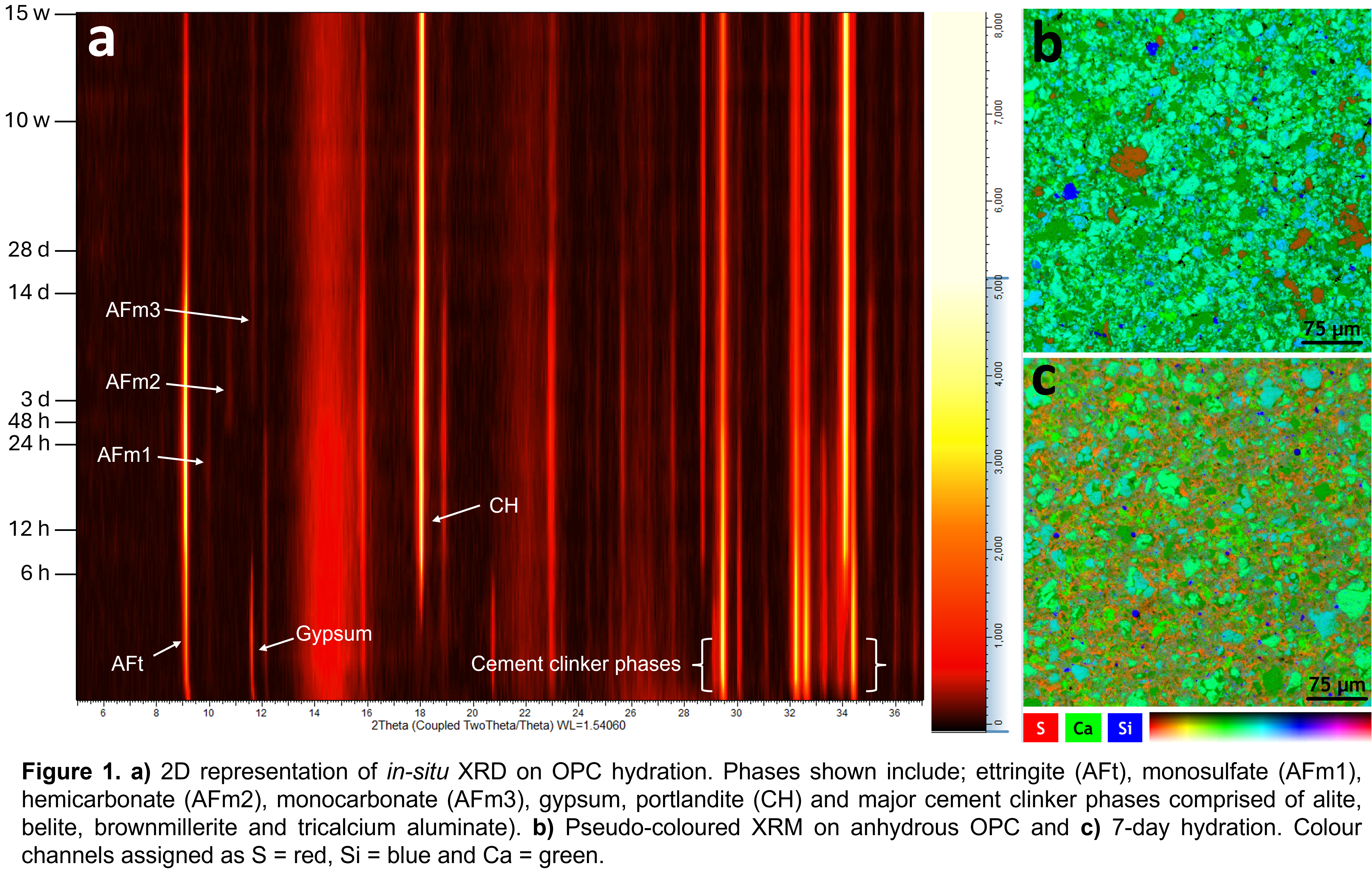
In-situ XRD results shown (Figure 1a), cover 15-weeks of OPC hydration. Induction and acceleration stages are noted by the consumption and conversion of gypsum to ettringite (AFt), in addition to the formation of portlandite (CH). Ettringite reaches a peak maximum at approximately 24 h, upon which conversion of ettringite to various hydrated calcium aluminate (AFm) phases begin. AFm1, monosulphate, is initially observed from 9 h with the formation of AFm2, hemicarbonate, occurring after this, reaching a peak maximum by 3 d. It is subsequently consumed and converted to AFm3, monocarbonate. AFm3 reaches a peak maximum at 7 days, after which it slowly begins to drop.
A supporting trend was observed in the XRM results, where a pellet of anhydrous OPC shows sulphur-rich regions attributed to gypsum (Figure 1b). These localised regions were not observed in the 7-day hydrated OPC mix, instead sulphur appears to be distributed throughout the sample matrix (Figure 1c), indicating ettringite (AFt) and AFm phase formation.
This work demonstrates the potential of using in-situ XRD with complementary microanalysis techniques for investigating cement hydration. These methods provide a pathway for studying the curing behaviour of PC-based systems and offer a framework for analysing more complex alternative cement formulations.
- Lord M. (2017). Rethinking Cement: Zero Carbon Industry Plan. Beyond Zero Emissions.
- Scrivener, K.L., John, V.M., Gartner, E.M. (2018). Cem. Concr. Res., 114, 2.
- Fanna, D.J., George, L., Moran, K., Wuhrer, R. (2024). Microscopy and Microanalysis, 30, 1.
- Wuhrer, R., Moran, K., Moran, L. (2006). Materials Forum, 30, 63.
- The authors acknowledge the AMCF at Western Sydney University for instrument access. The authors also thank Metrologi Pty Ltd., in particular, Scott Cheevers and Rhys Wuhrer for custom fabrication of components essential for in-situ analysis.