Oral Presentation Crystal36-AXAA Conference 2026
X-ray multimodal imaging from speckle-based to dual-sample imaging (143241)
For over a century, X-rays have been used to reveal the internal structure of opaque objects, driving innovations in healthcare, industry, and scientific research. However, conventional absorption-based X-ray imaging often struggles to distinguish materials with similar densities and cannot reveal unresolved micro- and nano-structural features below the resolution of the imaging system. To overcome these limitations, multimodal X-ray imaging methods have been developed that provide complementary phase-contrast and dark-field information in addition to conventional attenuation contrast. Whereas attenuation imaging measures the loss of X-ray intensity, phase contrast probes X-ray phase shifts introduced by the sample, leading to improved density resolution, and dark-field imaging is sensitive to small-angle scattering and unresolved refraction from sub-resolution structures.
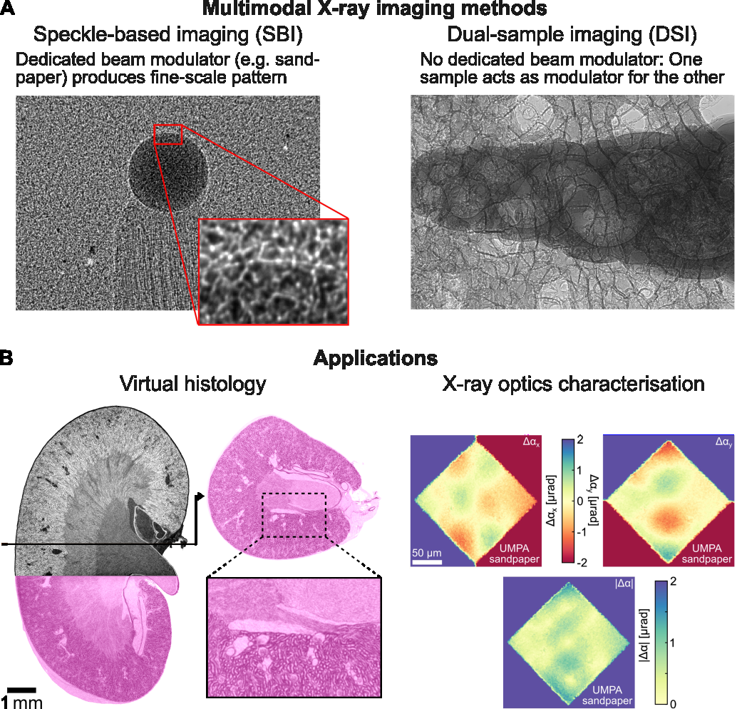
Most multimodal X-ray imaging approaches belong to the broader class of modulation-based imaging techniques, which use a structured illumination pattern to encode the sample’s phase and scattering information into the recorded detector signal. This modulation can be generated using optical elements in the beam, such as periodic gratings or grids, random diffusers like sandpaper, or other wavefront-marking structures, see Figure (A). Among these methods, X-ray speckle-based imaging (SBI), which uses a piece of sandpaper as a modulator, has emerged as a particularly versatile approach owing to its simple experimental implementation, high sensitivity, and compatibility with both synchrotron and laboratory sources [1-3]. The sample-induced local intensity reduction, displacement, and blurring of the speckle pattern can be analysed computationally, for example using the Unified Modulated Pattern Analysis (UMPA) framework [4], to quantitatively recover attenuation, phase, and dark-field signals, respectively. SBI and UMPA are now widely used at synchrotron facilities worldwide, including the Australian Synchrotron, Diamond (UK), ESRF (France), Elettra (Italy), and DESY (Germany). They have been demonstrated to benefit applications ranging from biomedical imaging to materials characterisation, geology, and X-ray metrology.
This presentation will provide an overview of the state-of-the-art and recent developments in SBI and showcase applications including virtual histology [5] and X-ray optics characterisation [6], see Figure (B).
We will furthermore discuss how viewing SBI and modulation-based imaging from a broader perspective naturally leads to new computational imaging concepts. In existing modulation-based imaging, information about the modulator is largely discarded after sample reconstruction, despite potentially carrying useful information. Furthermore, the ability to perform modulation-based imaging with a surprisingly diverse range of modulators—from precision-fabricated optics to simple pieces of sandpaper—suggests that the specific nature of the modulator is of secondary importance. Together, these observations motivate a broader view of modulation-based imaging in which the distinction between sample and optical element becomes increasingly blurred.
Building on this idea, we recently introduced dual-sample multimodal imaging (DSI), which generalises the concept of modulation-based imaging by using two samples in the beam as mutual modulators [7]. This enables spatial separation and multimodal image extraction of the two objects overlapping in projection from a single scan, effectively doubling imaging throughput without additional optical elements, while maintaining image quality comparable to SBI. DSI could open new opportunities for high-throughput inspection and constrained-environment imaging in research and industrial applications.
- [1] K. S. Morgan et al., Appl. Phys. Lett. 100, 124102 (2012).
- [2] S. Berujon et al., Phys. Rev. Lett. 108, 158102 (2012).
- [3] M.-C. Zdora, X-ray Phase-Contrast Imaging Using Near-Field Speckles (Springer, 2021).
- [4] M.-C. Zdora et al., Phys. Rev. Lett. 118, 203903 (2017).
- [5] M.-C. Zdora et al., Optica 7, 1221-1227 (2020).
- [6] M.-C. Zdora et al., Opt. Express 26, 4989-5004 (2018).
- [7] M.-C. Zdora et al., Optica, in press (2026).