Poster Presentation Crystal36-AXAA Conference 2026
Investigating the mechanism of proton delivery in cytochrome P450 enzymes using variable-temperature X-ray crystallography (145053)
Cytochrome P450 (CYP) monooxygenases are a family of heme-thiolate enzymes renowned for their ability to hydroxylate organic molecules regio- and enantio-selectively. Additionally, these enzymes catalyse a wealth of other reactions; these include C-C bond cleavage, desaturation, sulfoxidation, N,O,S-dealkylation and epoxidation. In humans, these enzymes have crucial roles (e.g. metabolising drugs). Because of their ability to carry out oxidation reactions with exceptional selectivity, there is immense interest in using these enzymes as catalysts to synthesise useful molecules (e.g. pharmaceuticals, drug metabolites, and flavour and fragrance compounds).
The active oxidant in most CYP reactions is a ferryl-oxo porphyrin radical cation species, referred to as Compound I. To generate the active species, the enzyme requires O2 from the atmosphere, as well as two electrons (from nicotinamide cofactors) and two protons (sourced from the surrounding solvent). Two highly conserved residues in the O2-binding groove above the heme are known to be essential for proper delivery of these protons. These conserved residues (commonly an aspartate/glutamate and a threonine) are known as the “acid-alcohol pair”. For example, in the bacterial enzymes P450cam (CYP101A1) and CYP199A4, the acid-alcohol pair consists of an aspartate (D251; the “acid”) and a threonine (T252; the “alcohol”).[1,2] Replacing the acidic D251 residue with a neutral asparagine (N) residue severely impairs the enzyme’s activity (resulting in a 100-fold reduction in activity), underscoring its importance. [1,2] This is due to slowing of the proton transfer steps. However, the exact mechanism of proton delivery in CYP enzymes has not been established.
In this study, we used variable-temperature X-ray crystallography to investigate the role of the acidic D251 residue in the model enzyme CYP199A4. Conventional protein X-ray crystallography is performed at 100 K (-173.15 °C), not room temperature, because cryogenic temperatures minimise radiation damage.[3-5] However, the standard practice of collecting data at cryogenic temperatures is not ideal.[3,4] This is because higher temperatures can be needed to view higher-energy protein conformations that provide insight into an enzyme’s catalytic mechanism, which are otherwise concealed at low temperatures.[3,4]
By raising the temperature of crystals of substrate-bound CYP199A4 from 100 K to 200 K in 50 K increments, we observed motion of the D251 side chain between two positions: one pointing into the active site, and one conformation pointing out of the active site into the solvent channel (Figure 1).[6] We found that the D251 side chain has a higher degree of mobility compared to other active-site residues.[6] Its ability to rotate in and out of the active site is consistent with a role in transferring protons from external solvent into the active site.[6] By performing molecular dynamics simulations, we identified a plausible proton relay involving residues E153, D251, R391 and E400.[6]
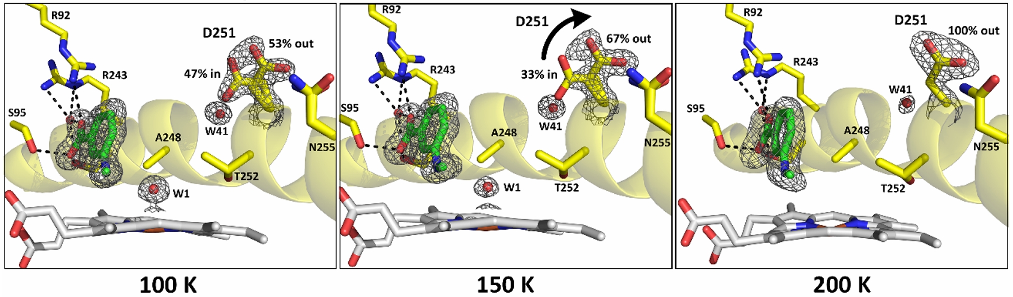
Figure 1. Variable-temperature X-ray crystallography was used to detect movement of the D251 side chain in and out of the active site in a crystal of ligand-bound CYP199A4.
- Nagano, S.; Poulos, T. L. Crystallographic Study on the Dioxygen Complex of Wild-type and Mutant Cytochrome P450cam: IMPLICATIONS FOR THE DIOXYGEN ACTIVATION MECHANISM *. Journal of Biological Chemistry 2005, 280 (36), 31659-31663. DOI: 10.1074/jbc.M505261200 (accessed 2026/07/26).
- Coleman, T.; Stok, J. E.; Podgorski, M. N.; Bruning, J. B.; De Voss, J. J.; Bell, S. G. Structural insights into the role of the acid-alcohol pair of residues required for dioxygen activation in cytochrome P450 enzymes. JBIC Journal of Biological Inorganic Chemistry 2020, 25 (4), 583-596. DOI: 10.1007/s00775-020-01781-4.
- Stachowski, T. R.; Fischer, M. Unfreezing structural biology for drug discovery. Nature Chemical Biology 2026, 22 (2), 169-179. DOI: 10.1038/s41589-025-02047-3.
- Fraser, J. S.; van den Bedem, H.; Samelson, A. J.; Lang, P. T.; Holton, J. M.; Echols, N.; Alber, T. Accessing protein conformational ensembles using room-temperature X-ray crystallography. Proceedings of the National Academy of Sciences 2011, 108 (39), 16247-16252. DOI: 10.1073/pnas.1111325108 (accessed 2026/07/26).
- Shelley, K. L.; Garman, E. F. Quantifying and comparing radiation damage in the Protein Data Bank. Nature Communications 2022, 13 (1), 1314. DOI: 10.1038/s41467-022-28934-0.
- Podgorski, M. N.; McDougal, D. P.; Campbell, E. C.; Bruning, J. B.; Bell, S. G. Structural basis for substrate-dependent allostery in oxygen activation by a cytochrome P450 enzyme revealed by analysis at different temperatures. Chemical Science 2026, 17 (15), 7742-7755. DOI: 10.1039/d5sc07539d (accessed 7/23/2026).