Poster Presentation Crystal36-AXAA Conference 2026
Tailoring AgTaO3 photocatalysts through La doping and surface-deposited Ag nanoparticles for enhanced PFAS degradation (145068)
Per- and polyfluoroalkyl substances (PFAS) are persistent environmental contaminants with exceptional chemical stability that makes them resistant to conventional treatment technologies. Semiconductor photocatalysis has emerged as a promising strategy for PFAS destruction, with catalyst structure and defect chemistry playing critical roles in determining photocatalytic performance. Here, we report the synthesis and comprehensive structural characterisation of La-doped AgTaO₃ photocatalysts, developed for the mineralisation of perfluorooctanesulfonic acid (PFOS).
La-doped AgTaO₃ photocatalysts were prepared via a high-temperature solid-state synthesis route. Powder X-ray diffraction (PXRD) confirmed the formation of trigonal-phase AgTaO₃ and UV–Visible diffuse reflectance spectroscopy (UV-Vis DRS) was used to calculate the bandgap of the materials (3.4 eV, 365 nm). Scanning electron microscopy coupled with energy-dispersive X-ray spectroscopy (SEM-EDS) and high-resolution scanning transmission electron microscopy (HR-STEM) revealed the AgTaO₃ nanoparticles were approximately 100–200 nm in size and decorated with smaller Ag nanoparticles (7–15 nm) at the surface (Figure 1). Variable-wavelength X-ray photoelectron spectroscopy (XPS) at the Australian Synchrotron was further employed to investigate changes in surface and near-surface defect chemistry associated with La incorporation and Ag deposition.
Continuous flow photocatalysis under 365 nm irradiation achieved > 80% mineralisation of PFOS, demonstrating the effectiveness of the photocatalysts for degrading highly persistent, fluorinated contaminants. Correlating advanced X-ray characterisation with photocatalytic performance provides new insight into the roles of La doping, surface-deposited Ag nanoparticles, and lattice defects in enhancing PFAS mineralisation. This work demonstrates how detailed structural and defect analysis can guide the rational design of photocatalysts for environmental remediation applications.
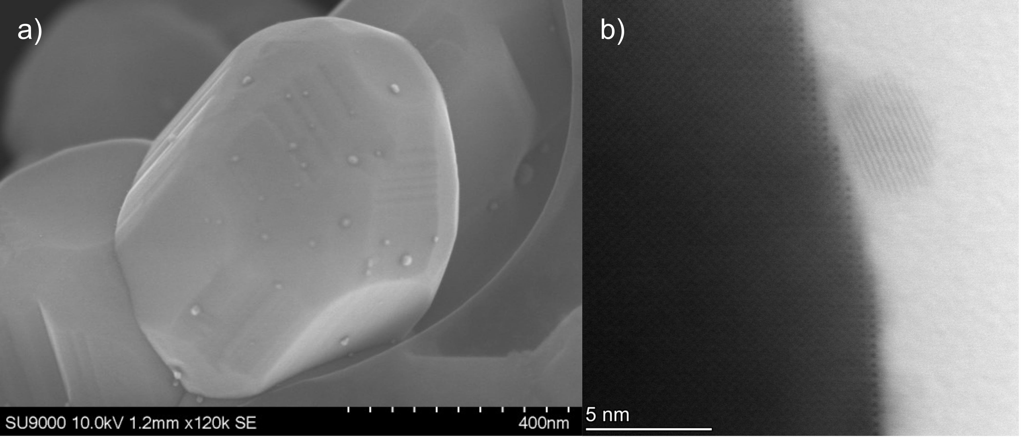
Figure 1: (a) SEM image (b) high-angle annular dark field image from HR-STEM analysis highlighting Ag nanoparticles deposited on the surface of La-doped AgTaO3 nanoparticles.
- Kato, H.; Kobayashi, H.; Kudo, A. Role of Ag+ in the band structures and photocatalytic properties of AgMO3 (M: Ta and Nb) with the perovskite structure. The Journal of Physical Chemistry B 2002, 106 (48), 12441-12447.
- Matthews, R. K.; Nguyen, H. T.; Yang, F.; Pukala, T. L.; Evans, J. D.; Shearer, C. J. La doping ATaO₃ (A = Li, Na, K) to improve performance for photocatalytic pollutant degradation. Chemistry of Materials 2025, 37 (10), 3696-3708.
- Matthews, R. K.; Day, M. L.; Rahman, T. M.; Dadkhah, M.; Toyota, Y.; Kobayashi, Y.; Shearer, C. J. Development of a Continuous Flow Photoreactor for Photocatalytic Pollutant Degradation. Colloids and Surfaces A: Physicochemical and Engineering Aspects 2025, 735, 139354.